
 Diagnosing Heart Disease With a Chest X-Ray
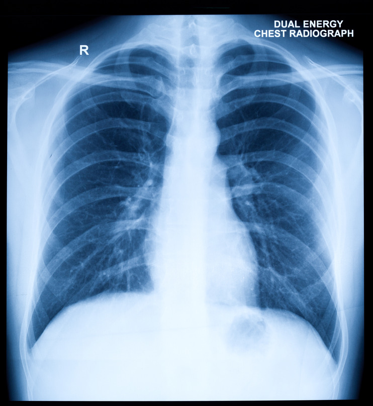
A chest X-ray (also called chest film) uses a very small amount of radiation to produce an image of the heart, lungs, and chest bones on film. A physician may use a chest X-ray to:
- Look at the structures of the chest (bones, heart, lungs).
- Evaluate placement of devices (pacemakers, defibrillators) or tubes placed during hospitalization for treatment and monitoring (catheters, chest tubes).
- Diagnose lung and cardiac diseases.
What Happens During a Chest X-Ray?
A chest X-ray can be performed in our office. You will be asked to remove all clothes and metallic jewelry from the waist up and put on a hospital gown for the test.
If you are able, you will be asked to stand very still with your chest against the cassette that contains the digital film. The X-ray machine will then send a beam of x-rays through your chest onto the film to create a picture. Bones and other dense areas show up as lighter shades of gray while areas that don’t absorb the radiation appear as dark gray.
You will be asked to hold your breath for a few seconds to generate better images. Then you will be asked to do the same thing, but with your left side against the cassette and your arms elevated.
The entire test takes no more than 5 to 10 minutes.