
 Transesophageal Echocardiography (TEE) is a type of echocardiography (echo). Echo shows the size and shape of the heart and how well the heart chambers and valves are working.
Echo can pinpoint areas of heart muscle that aren’t contracting well because of poor blood flow or injury from a previous heart attack.
Echo also can detect possible blood clots inside the heart, fluid buildup in the pericardium (the sac around the heart), and problems with the aorta. The aorta is the main artery that carries oxygen-rich blood from your heart to your body.
Overview
During echo, a device called a transducer is used to send sound waves (called ultrasound) to the heart. As the ultrasound waves bounce off the structures of the heart, a computer in the echo machine converts them into pictures on a screen.
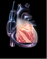
TEE involves a flexible tube (probe) with a transducer at its tip. Your doctor will guide the probe down your throat and into your esophagus (the passage leading from your mouth to your stomach). This approach allows your doctor to get more detailed pictures of your heart because the esophagus is directly behind the heart.
TEE can help doctors diagnose heart and blood vessel diseases and conditions in adults and children. Doctors also may use TEE to guide cardiac catheterization (KATH-eh-ter-ih-ZA-shun), help prepare for surgery, or assess a patient’s status during or after surgery.
Doctors may use TEE in addition to transthoracic (tranz-thor-AS-ik) echo (TTE), the most common type of echo. If TTE pictures don’t give doctors enough information, they may recommend TEE to get more detailed pictures.
Outlook
TEE has a low risk of complications in both adults and children. Even newborns can have TEE.
Article Courtesy of NIH